Home
/ Animal Cell Structure Label Figure 4.2 : 4 2 Parts Of A Eukaryotic Cell / 2.3.2 annotate the diagram from 2.3.1 with the functions of each named structure.
Animal Cell Structure Label Figure 4.2 : 4 2 Parts Of A Eukaryotic Cell / 2.3.2 annotate the diagram from 2.3.1 with the functions of each named structure.
Animal Cell Structure Label Figure 4.2 : 4 2 Parts Of A Eukaryotic Cell / 2.3.2 annotate the diagram from 2.3.1 with the functions of each named structure.. The internal structure of a chloroplast, with a granal stack of thylakoids circled. It shows various stages of mitosis in an animal cell. These figures represent a eukaryotic cell and a prokaryotic cell. Last updated on wed, 16 dec 2020 | medical terminology. Ological concepts animal cell structure label figure 4.2.
These will be the focus of this these will be further discussed in the photosynthesis concept. Ribosomes the site of protein building this is where translation takes place mrna in language the worksheets recommended for 7th and 8th grade students feature labeled animal and plant cell structure charts and cross section charts cell. Cells are important elements of living. See how a generalized structure of an animal cell and plant cell look with labeled diagrams. Figure 4.2a the sizes of living things and their components.
Cell Biology Wikipedia from upload.wikimedia.org Cell organelles structure and parts. 3 structures outside the cell membrane. All prokaryotes have chromosomal dna localized in a nucleoid the plant cell has a cell wall, chloroplasts, plastids, and a central vacuole—structures not in animal cells. Figure 4.8 these figures show the major organelles and other cell components of (a) a typical animal cell and (b) a typical eukaryotic plant cell. Therefore, detailed neuronal morphology is required to understand normal neuronal function and pathological mechanisms, such as those that occur in autism. Naturally, if they are the structural and functional unit of living, there has to be something peculiar about them? The animal cell is made up of several structural organelles enclosed in the plasma membrane, that enable it to function properly, eliciting mechanisms. Different cell types of neurons form complicated circuits in the brain.
Animal cell anatomy on white background.
Figure 4.5 this figure shows the generalized structure of a prokaryotic cell. Be it humans or plants or animals, every living organism is composed of cells. Label a diagram of a typical cell. Figure 4.20b animal cells are joined by three different types of junctions. These figures represent a eukaryotic cell and a prokaryotic cell. The internal structure of a chloroplast, with a granal stack of thylakoids circled. Learn about the size and function of plant and animal cells for gcse combined science, aqa. Which organelle contains its own dna? Cell is a tiny structure and functional unit of a living organism containing various parts known as organelles. Cell structure and function notes: While the plant cell resembles rectangular shape and. The structure of an animal cell, with labeled parts. What structures are found within the nucleus?
Animal cell definition with cell size and shape. While the plant cell resembles rectangular shape and. Pictures cells that have structures unlabled, students must write the labels in, this is intended for more advanced biology students. Diagram of animal cell, created with biorender.com. Animal cell functions are solely dependent on the organelles and structures associated with the cell.
Unique Characteristics Of Eukaryotic Cells Microbiology from s3-us-west-2.amazonaws.com Animal cells do not have these rigid exteriors. Diagram of animal cell, created with biorender.com. A single cell of a unicellular organism like an amoeba b. Animal cells are common names for eukaryotic cells that make up animal tissue. Learn the parts of animal and plant cells by labeling the diagrams. All living organisms are made up of cells that. The animal cell is made up of several structural organelles enclosed in the plasma membrane, that enable it to function properly, eliciting mechanisms. A plasma membrane encloses every cell including a.
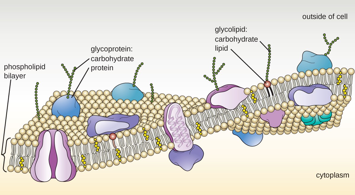
Cell membrane structure and function.
Animal cell definition with cell size and shape. A single cell of a unicellular organism like an amoeba b. Last updated on wed, 16 dec 2020 | medical terminology. Every animal cell has a cell membrane, cytoplasm, and a nucleus, but not all cells have every structure shown here. Form the basic unit of structure and function. You will get a picture of a animal or plant cell, label the cell. The animal cell is made up of several structural organelles enclosed in the plasma membrane, that enable it to function properly, eliciting mechanisms. Illustrated in figure 2 are a pair of fibroblast deer skin cells that have been labeled with fluorescent probes and photographed in the microscope to reveal. These will be the focus of this these will be further discussed in the photosynthesis concept. Animal cell and fungal yeast cell structure. (reprinted with permission from cohen bj, wood dl. Here, we developed a strategy to sparsely label the same type of neurons. These figures represent a eukaryotic cell and a prokaryotic cell.
Form the basic unit of structure and function. The structure and contents of a typical animal cell. The diameter of most plant and animal cells is about. In the labeled animal cell diagram, it is nearly circular in shape and lacks outer cell wall; Naturally, if they are the structural and functional unit of living, there has to be something peculiar about them?
A Tour Of The Cell View As Single Page from www.open.edu The central and rightmost cell are in interphase, so their dna is diffuse and the entire nuclei are labelled. Transcribed image text from this question. The structure and contents of a typical animal cell. Pictures cells that have structures unlabled, students must write the labels in, this is intended for more advanced biology students. There are other distinct differences between plant and animal cells. (reprinted with permission from cohen bj, wood dl. Draw and label the image of the cheek cells on the activity sheet b. It should be large, clear and with specific labels.
Animal cell definition with cell size and shape.
Figure 4.5 this figure shows the generalized structure of a prokaryotic cell. Ribosomes the site of protein building this is where translation takes place mrna in language the worksheets recommended for 7th and 8th grade students feature labeled animal and plant cell structure charts and cross section charts cell. Printable animal cell diagram labeled unlabeled and blank. Illustrated in figure 2 are a pair of fibroblast deer skin cells that have been labeled with fluorescent probes and photographed in the microscope to reveal. Diagram of animal cell, created with biorender.com. It shows various stages of mitosis in an animal cell. In the labeled animal cell diagram, it is nearly circular in shape and lacks outer cell wall; Label a diagram of a typical cell. All organelles seem to share many properties with bacteria. Different kinds of animals have different numbers of cells, but most have millions animal cells contain small structures called organelles, which help carry out the normal operations of a cell. It should be large, clear and with specific labels. 3 structures outside the cell membrane. Every animal cell has a cell membrane, cytoplasm, and a nucleus, but not all cells have every structure shown here.
Share :
Post a Comment
for "Animal Cell Structure Label Figure 4.2 : 4 2 Parts Of A Eukaryotic Cell / 2.3.2 annotate the diagram from 2.3.1 with the functions of each named structure."


Post a Comment for "Animal Cell Structure Label Figure 4.2 : 4 2 Parts Of A Eukaryotic Cell / 2.3.2 annotate the diagram from 2.3.1 with the functions of each named structure."