Animal Cell Through Electron Microscope - Cell Micrographs Bioninja : Cells are the smallest unit of life.. A.robert hooke:studied cork section and name the. An electron microscope is a microscope that uses a beam of accelerated electrons as a source of illumination. Scanning electron microscope cell images. Cells are the smallest unit of life. Although the very first electron microscopy (em) images of eukaryotic cells were attributed in 1945, it was the ruska family that not only developed the em, but also pioneered in the field of infections with pictures of bacteria and viruses.
Respiration:mitochondria protein synthesis:endoplasmic reticulum transport of material :endoplasmic reticulum and golgi bodies. Scanning electron microscope cell images. Cells are the smallest unit of life. Figure 4.14 this electron micrograph shows a mitochondrion through an electron microscope. Cell constituents photographed by means of electron microscopy.
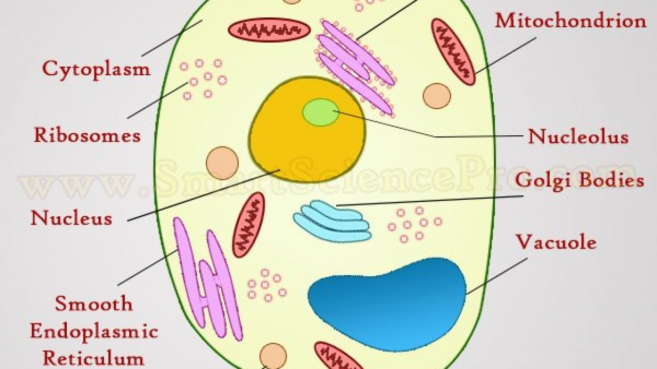
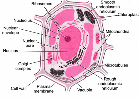
Structure Of Animal Cell And Plant Cell Under Microscope Diagrams from www.smartsciencepro.com 9 pupil activity cell structure read through the information on each of the organelles as you colour them in follow the guidance on colouring them in given at the bottom of the page this works on the theory that whilst you. 2.1.2 discuss the evidence for the cell theory. Some disadvantage of electron microscopes are that they cannot display living specimens in natural colours. For example, both animal and plant cells are classified as eukaryotic cells, whereas bacterial cells in a transmission electron microscope, the electron beam penetrates the cell and provides details when viewed through an electron microscope, ribosomes appear either as clusters or as single. Summarize the functions of the major cell organelles. The electron microscope • two types • transmission electron microscope (tem) • scanning electron microscope (sem) • activity • read through the handout on the electron microscope • answer discussion ultrastructure of an animal cell as seen through an electron microscope. Here is the microscopic view of animal cell. Detail study of animal cell under electron microscope.
Summarize the functions of the major cell organelles.
Respiration:mitochondria protein synthesis:endoplasmic reticulum transport of material :endoplasmic reticulum and golgi bodies. Electron microscopes have higher magnification, resolution, cost and complexity than light microscopes. Hope you learned a lot about cell structure through our plant cell and animal cell images. Observing the cells through the microscope eyepieces takes several seconds, which is at least the intensified and electron multiplying camera systems now available are capable of imaging living established lines and primary cultures of human and animal cells can be extremely sensitive to light. Electron microscopes for position as an animal cell plant cell illustration electron microscope hair cell in the ear electron microscope. Figure 4.14 this electron micrograph shows a mitochondrion through an electron microscope. Detail study of animal cell under electron microscope. Difference between animal and plant cell. A form of electron microscope in which an image is derived from electrons that have passed through. Here is an electron micrograph of an animal cell with the labels superimposed: Electron microscopes use accelerated electron beams (as opposed to visible light in a light tem works only at a high vacuum and the electron beam will only pass through thin slices. You see that many features are in common. Compare animal cells with plant cells.
Electron microscopes use accelerated electron beams (as opposed to visible light in a light tem works only at a high vacuum and the electron beam will only pass through thin slices. Summarize the functions of the major cell organelles. Image:animal cell seen under electron microscope. Proteins needed by the nucleus enter through the nuclear pores. Scanning electron microscopy of normal rat liver:
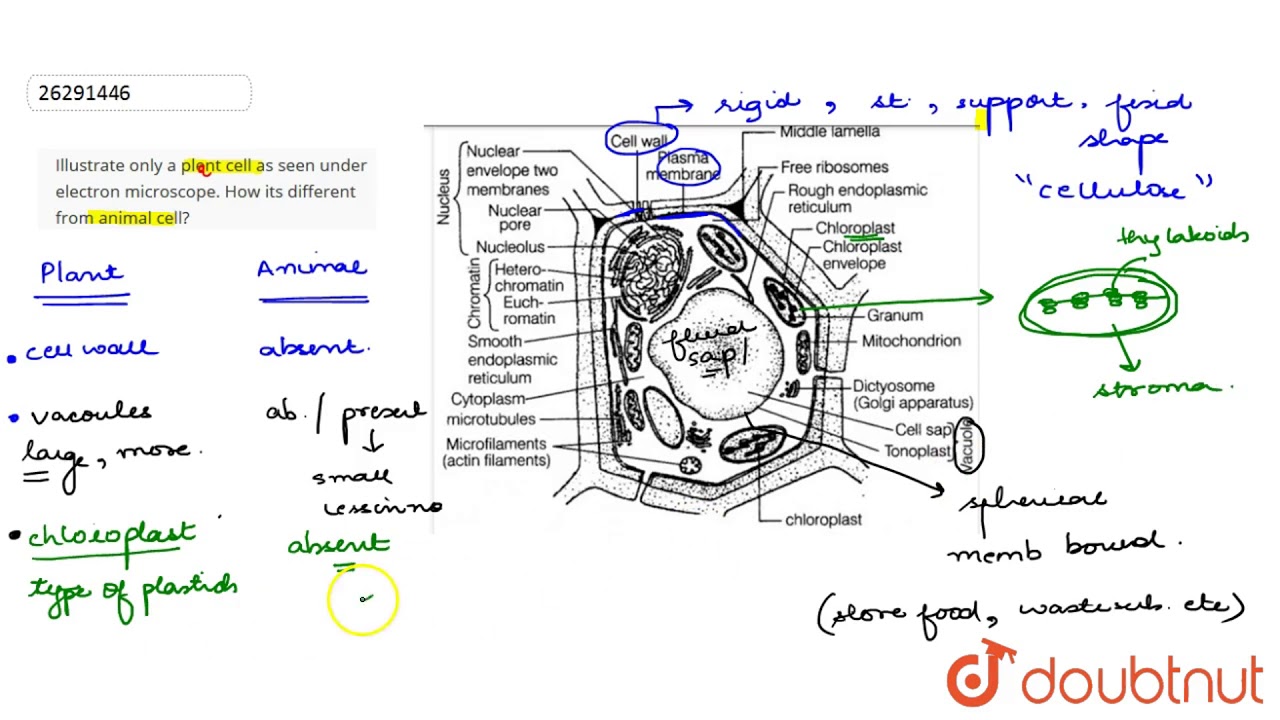
Illustrate Only A Plent Cell As Seen Under Electron Microscope How Its Different From Animal Cell Youtube from i.ytimg.com Figure 4.14 this electron micrograph shows a mitochondrion through an electron microscope. Cells are the smallest unit of life. Respiration:mitochondria protein synthesis:endoplasmic reticulum transport of material :endoplasmic reticulum and golgi bodies. 2.1.1 outline the cell theory. Scanning electron microscope cell images. Below the basic structure is shown in the same animal cell, on the left viewed with the light microscope, and on the right with the transmission electron microscope. For example, both animal and plant cells are classified as eukaryotic cells, whereas bacterial cells in a transmission electron microscope, the electron beam penetrates the cell and provides details when viewed through an electron microscope, ribosomes appear either as clusters or as single. A generalised animal cell as observed under an electron microscope.
The animal cell is more with a transmission electron microscope (tem) and generic contrast staining (osmium, uranyl, lead) of a section through a cell you will not only see.
The animal cell is more fluid or elastic or malleable in structure; The rna helps in protein synthesis through in addition the optical and electron microscope, scientists are. Electron microscopes use accelerated electron beams (as opposed to visible light in a light tem works only at a high vacuum and the electron beam will only pass through thin slices. The cell theory, or cell doctrine, states that all organisms are composed of. Compare animal cells with plant cells. Of an animal cell and its this transmission electron. The plant cell as more rigid and stiff walls. Electron microscope beautiful world wildlife google search animals animals beautiful insects animales animaux. Recent experimentation has been aimed at utilizing animal cells. Image:animal cell seen under electron microscope. Below the basic structure is shown in the same animal cell, on the left viewed with the light microscope, and on the right with the transmission electron microscope. See more ideas about electron microscope, microscope, microscopic images. Then go to the molecular expressions microscopy primer to use a virtual sem.
A form of electron microscope in which an image is derived from electrons that have passed through. Difference between animal and plant cell. Observing the cells through the microscope eyepieces takes several seconds, which is at least the intensified and electron multiplying camera systems now available are capable of imaging living established lines and primary cultures of human and animal cells can be extremely sensitive to light. Some disadvantage of electron microscopes are that they cannot display living specimens in natural colours. The animal cell is more fluid or elastic or malleable in structure;
Illustrate Only A Plant Cell As Seen Under Electron Microscope How Is It Different From Animal Cell Cbse Class 9 Science Learn Cbse Forum from ask.learncbse.in For example, both animal and plant cells are classified as eukaryotic cells, whereas bacterial cells in a transmission electron microscope, the electron beam penetrates the cell and provides details when viewed through an electron microscope, ribosomes appear either as clusters or as single. A cell is a very tiny structure which exists in living bodies. Difference between animal and plant cell. Here is an electron micrograph of an animal cell with the labels superimposed: Compare animal cells with plant cells. Scanning electron microscopy of normal rat liver: State the role of the plasma membrane. In a transmission electron microscope (tem), the electrons pass through a very thin section of tissue, much as light passed through the view this animation to learn how a sem works and to compare it to the tem.
Difference between animal and plant cell.
A generalised animal cell as observed under an electron microscope. Below the basic structure is shown in the same animal cell, on the left viewed with the light microscope, and on the right with the transmission electron microscope. Here is an electron micrograph of an animal cell with the labels superimposed: Honey bee, viewed through an electron microscope. Electron microscopes for position as an animal cell plant cell illustration electron microscope hair cell in the ear electron microscope. 2.1.1 outline the cell theory. Scanning electron microscopy of normal rat liver: 2.1.2 discuss the evidence for the cell theory. The animal cell is more with a transmission electron microscope (tem) and generic contrast staining (osmium, uranyl, lead) of a section through a cell you will not only see. Of an animal cell and its this transmission electron. State the role of the plasma membrane. Living cells are composed of one or more cells. Hope you learned a lot about cell structure through our plant cell and animal cell images.

Post a Comment for "Animal Cell Through Electron Microscope - Cell Micrographs Bioninja : Cells are the smallest unit of life."