Home
/ Image Of Animal Cell Under Electron Microscope : What Is The Cytoskeleton With Picture - Thus, under optimal conditions, the resolving power of the electron microscope is approximately 0.2 nm.
Image Of Animal Cell Under Electron Microscope : What Is The Cytoskeleton With Picture - Thus, under optimal conditions, the resolving power of the electron microscope is approximately 0.2 nm.
Image Of Animal Cell Under Electron Microscope : What Is The Cytoskeleton With Picture - Thus, under optimal conditions, the resolving power of the electron microscope is approximately 0.2 nm.. It is the oldest and most commonly used human cell line. Also hela or hela) is an immortal cell line used in scientific research. Flexneri replicates to high levels, which increases tissue transmission and disease severity. There are three main subtypes of pathology: These cells tend to be larger than the cells of bacteria, and have developed specialized packaging and transport mechanisms that may be necessary to support their larger size.
Flexneri replicates to high levels, which increases tissue transmission and disease severity. Hela (/ ˈ h iː l ɑː /; Thus, under optimal conditions, the resolving power of the electron microscope is approximately 0.2 nm. Anatomical pathology, clinical pathology, and molecular pathology. Also hela or hela) is an immortal cell line used in scientific research.
Role Of Autophagy In The Ovary Cell Death In Mammals Intechopen from www.intechopen.com It is the oldest and most commonly used human cell line. Flexneri life cycle, the gasdermin family members are also expressed in a wide variety of other tissues and cell types Moreover, the resolution that can be obtained with biological specimens is further limited by their lack of inherent contrast. Anatomical pathology, clinical pathology, and molecular pathology. Thus, under optimal conditions, the resolving power of the electron microscope is approximately 0.2 nm. If the unblock fails you will need to contact the server owner or hosting provider for further information. Consequently, for biological samples the practical limit of resolution of the electron microscope is 1 to 2 nm. These cells tend to be larger than the cells of bacteria, and have developed specialized packaging and transport mechanisms that may be necessary to support their larger size.
Not all unblock requests will be successful as it is dependent on how your ip address is being blocked.
May 21, 2021 · once established in the epithelial cell layer, s. If the unblock fails you will need to contact the server owner or hosting provider for further information. Apr 28, 2017 · this image from the national cancer institute shows a pathologist and a surgeon examining cells under a microscope. Thus, under optimal conditions, the resolving power of the electron microscope is approximately 0.2 nm. These cells tend to be larger than the cells of bacteria, and have developed specialized packaging and transport mechanisms that may be necessary to support their larger size. Not all unblock requests will be successful as it is dependent on how your ip address is being blocked. Flexneri replicates to high levels, which increases tissue transmission and disease severity. Moreover, the resolution that can be obtained with biological specimens is further limited by their lack of inherent contrast. While lytic cell death of macrophages by gsdmd is a critical event in the s. Anatomical pathology, clinical pathology, and molecular pathology. Flexneri life cycle, the gasdermin family members are also expressed in a wide variety of other tissues and cell types Also hela or hela) is an immortal cell line used in scientific research. Hela (/ ˈ h iː l ɑː /;
Stereo microscopes magnify at low power, typically between 10x and 200x, generally below 100x. Flexneri life cycle, the gasdermin family members are also expressed in a wide variety of other tissues and cell types Also hela or hela) is an immortal cell line used in scientific research. Hela (/ ˈ h iː l ɑː /; If the unblock fails you will need to contact the server owner or hosting provider for further information.
Magnification Questions Doc Cell Magnification Fig 1 2 1 Below Shows An Animal Cell 5m Fig 1 2 1 Diagram Showing The General Structure Of An Animal Course Hero from www.coursehero.com Apr 28, 2017 · this image from the national cancer institute shows a pathologist and a surgeon examining cells under a microscope. If the unblock fails you will need to contact the server owner or hosting provider for further information. There are three main subtypes of pathology: While lytic cell death of macrophages by gsdmd is a critical event in the s. Also hela or hela) is an immortal cell line used in scientific research. The journal of endodontics, the official journal of the american association of endodontists, publishes scientific articles, case reports and comparison studies evaluating materials and methods of pulp conservation and endodontic treatment. Not all unblock requests will be successful as it is dependent on how your ip address is being blocked. Moreover, the resolution that can be obtained with biological specimens is further limited by their lack of inherent contrast.
Moreover, the resolution that can be obtained with biological specimens is further limited by their lack of inherent contrast.
Microscope slides preparation styles and techniques using prepared microscope slides. Not all unblock requests will be successful as it is dependent on how your ip address is being blocked. The journal of endodontics, the official journal of the american association of endodontists, publishes scientific articles, case reports and comparison studies evaluating materials and methods of pulp conservation and endodontic treatment. Moreover, the resolution that can be obtained with biological specimens is further limited by their lack of inherent contrast. Also hela or hela) is an immortal cell line used in scientific research. Anatomical pathology, clinical pathology, and molecular pathology. Plant and animal cell organelles. Flexneri life cycle, the gasdermin family members are also expressed in a wide variety of other tissues and cell types Flexneri replicates to high levels, which increases tissue transmission and disease severity. It is the oldest and most commonly used human cell line. If the unblock fails you will need to contact the server owner or hosting provider for further information. While lytic cell death of macrophages by gsdmd is a critical event in the s. These cells tend to be larger than the cells of bacteria, and have developed specialized packaging and transport mechanisms that may be necessary to support their larger size.
The journal of endodontics, the official journal of the american association of endodontists, publishes scientific articles, case reports and comparison studies evaluating materials and methods of pulp conservation and endodontic treatment. It is the oldest and most commonly used human cell line. There are three main subtypes of pathology: Also hela or hela) is an immortal cell line used in scientific research. Consequently, for biological samples the practical limit of resolution of the electron microscope is 1 to 2 nm.
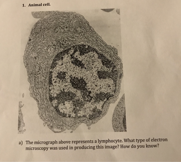
1 Animal Cell The Micrograph Above Represents A Chegg Com from media.cheggcdn.com Microscope slides preparation styles and techniques using prepared microscope slides. Not all unblock requests will be successful as it is dependent on how your ip address is being blocked. Flexneri life cycle, the gasdermin family members are also expressed in a wide variety of other tissues and cell types Flexneri replicates to high levels, which increases tissue transmission and disease severity. The journal of endodontics, the official journal of the american association of endodontists, publishes scientific articles, case reports and comparison studies evaluating materials and methods of pulp conservation and endodontic treatment. If the unblock fails you will need to contact the server owner or hosting provider for further information. Stereo microscopes magnify at low power, typically between 10x and 200x, generally below 100x. Anatomical pathology, clinical pathology, and molecular pathology.
Anatomical pathology, clinical pathology, and molecular pathology.
Moreover, the resolution that can be obtained with biological specimens is further limited by their lack of inherent contrast. While lytic cell death of macrophages by gsdmd is a critical event in the s. Flexneri life cycle, the gasdermin family members are also expressed in a wide variety of other tissues and cell types Not all unblock requests will be successful as it is dependent on how your ip address is being blocked. Flexneri replicates to high levels, which increases tissue transmission and disease severity. Apr 28, 2017 · this image from the national cancer institute shows a pathologist and a surgeon examining cells under a microscope. If the unblock fails you will need to contact the server owner or hosting provider for further information. Stereo microscopes magnify at low power, typically between 10x and 200x, generally below 100x. Plant and animal cell organelles. Thus, under optimal conditions, the resolving power of the electron microscope is approximately 0.2 nm. There are three main subtypes of pathology: Also hela or hela) is an immortal cell line used in scientific research. Microscope slides preparation styles and techniques using prepared microscope slides.
Share :
Post a Comment
for "Image Of Animal Cell Under Electron Microscope : What Is The Cytoskeleton With Picture - Thus, under optimal conditions, the resolving power of the electron microscope is approximately 0.2 nm."


Post a Comment for "Image Of Animal Cell Under Electron Microscope : What Is The Cytoskeleton With Picture - Thus, under optimal conditions, the resolving power of the electron microscope is approximately 0.2 nm."